
 Thomas Höllt
Vis Group, Department of Informatics, University of Bergen, Norway
From Cellular Composition to Tissue Organization: Visual Analytics for Single-Cell and Spatial Data
Abstract • Single-cell and spatially resolved molecular profiling technologies, such as mass cytometry and transcriptomics, are enabling increasingly detailed investigations of cellular heterogeneity and tissue organization. However, analyzing these high-dimensional datasets remains challenging, as they may comprise hundreds of thousands of observations, each characterized by tens to thousands of measured features.
Visual analytics (VA) and dimensionality reduction (DR) have proven valuable for the exploratory analysis of such data. In this talk, I will discuss VA methods and applications ranging from the analysis of cell-type composition in large samples to the investigation of cell–cell interactions and tissue organization in spatially resolved data. We will examine how high-performance DR techniques enable interactive workflows and scale to large, high-dimensional datasets, both non-spatial and spatially resolved.
Bio • Thomas is a professor for visualization in the Vis Group at the University of Bergen, Norway. Before that, he was faculty member at TU Delft and the Leiden University Medical Center. Thomas obtained his PhD from King Abdullah University of Science and Technology in 2013.
His research interests include Visualization and Visual Analytics, with a focus on high-dimensional data and bio-/medical applications. He initiated the research and development of Cytosplore, an interactive visual analytics platform for cytometry data which is used by researchers world-wide.
 Rok Romih
Institute of Cell Biology, Faculty of Medicine, University of Ljubljana, Slovenia
Visualisation of Multimodal Microscopy Datasets for Resolving Urinary Bladder Function
Abstract • The urinary bladder is a hollow muscular organ located at the base of the pelvis that stores urine. Its primary function is to withstand repeated cycles of gradual distension and rapid contraction while maintaining an impermeable barrier between urine and blood. The structure of the bladder wall, consisting of three main tissue layers, is well adapted to these functions. The luminal surface is lined by a specialized epithelium, the urothelium. Beneath it lies the lamina propria, composed of connective tissue, and the outer layer consists of smooth muscle cells forming the detrusor muscle. The urothelium is composed of undifferentiated basal cells, intermediate cells, and terminally differentiated superficial (umbrella) cells. A unique feature of umbrella cells is the presence of urothelial plaques formed by highly organized transmembrane proteins known as uroplakins. These plaques are found both in fusiform vesicles and in the apical plasma membrane, where they form a permeability barrier. Disruption of this blood–urine permeability barrier is associated with various bladder-related diseases, such as interstitial cystitis/bladder pain syndrome. Despite two-dimensional microscopic observations of the urinary bladder wall dating back to the 17th century, important questions regarding urothelial structure–function relationships remain unresolved. Recent advances in three-dimensional microscopy, combined with modern visualization techniques, offer new opportunities to address these questions. In the presentation, I will describe approaches for acquiring 3D datasets of bladder samples at different resolution limits, as well as methods for their visualization. The integration of these data may ultimately provide new insights into bladder function under both physiological and pathological conditions.
Bio • Rok Romih is a Full Professor at the Institute of Cell Biology, Faculty of Medicine, University of Ljubljana, Slovenia. His doctoral research focused on the differentiation of urothelial cells and the regeneration of urinary bladder urothelium following injury. His research encompasses the development of advanced methods for biological sample preparation for electron microscopy, ultrastructural protein localization, vesicular trafficking, and intercellular signaling mechanisms. He is actively involved in undergraduate and postgraduate education, teaching cell biology for medical, dental medical, biochemistry, and biomedicine students. As Head of the Centre for Electron Microscopy, he leads research activities in advanced imaging and microscopy while fostering numerous national and international scientific collaborations.
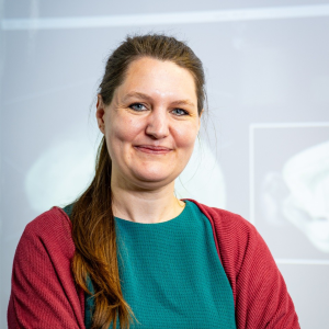 Sophia Ulonska
Vienna Research Center for Visual Computing, Austria
Interactive Visual Computing Tools for Multi-Modal Brain Data Exploration
Abstract • Understanding the brain in all its complexity is essential not only for scientific curiosity but also for medical, social, and economic reasons. In Europe alone, brain disorders affected 179 million people with annual costs estimated at €800 billion in 2010 [1]. While advances like spatial transcriptomics and AI enable new discoveries, neuroscience remains labor- and resource-intensive, often involving animal experiments and taxpayer funding. This demands careful planning and maximum utilization of existing data.
Connecting and interpreting diverse datasets is technically complex. Neuroscience data spans multiple species (e.g., fruit flies, mice, or humans), modalities (gene expression, connectivity, activity, …) and heterogeneous spatial scales from micro-scale microscopy data to meso-scale fMRI data. Integrating these data types while retaining spatial and anatomical context requires high computational expertise, which many neuroscientists lack. Interactive visual computing tools aim to support them in this task.
In this talk, besides a short overview of tools from various initiatives (Allen Institute, EBRAINS, or NeuronBridge), two complementary interactive visual computing platforms developed at VRVis GmbH will be presented: BrainTrawler provides free, public access to multi-modal brain datasets from humans and mice, integrating spatial genetic, connectomic, and functional data across scales. It features an innovative method for deriving circuits from brain activity signals in real time, with the capability to mine differentially expressed genes in the entire circuit. LarvalBrain offers unprecedented interactive access to Drosophila larva data across developmental stages within a standardized atlas framework and allows researchers to inform future behavioral experiments by interactive queries. In summary, these visual computing tools are valuable for supporting neuroscientists in investigating the brain.
[1] M. DiLuca and J. Olesen, “The Cost of Brain Diseases: A Burden or a Challenge?,” Neuron, vol. 82, no. 6, pp. 1205–1208, June 2014, doi: 10.1016/j.neuron.2014.05.044.
Bio • Dr Sophia Ulonska is a senior researcher at the nonprofit research center VRVis GmbH, located in Vienna, Austria. She is a mathematician who obtained her PhD in Technical Science in the field of Chemical and Process Engineering at TU Wien by developing algorithms to model, monitor, and control bacterial and mammalian cell fermentation processes. At VRVis, she works on computational neuroscience, particularly in areas such as multimodal data analysis, neural decoding and evolution of the brain. Furthermore, she develops scientific visual computing software that allows neuroscientists to interactively explore multimodal brain data in humans, mice, and Drosophila melanogaster.